Home

Research Areas
This field involves developing algorithms and computational methods to analyze and interpret medical images, such as MRI, CT scans, and X-rays. The goal is to improve disease diagnosis, treatment planning, and monitoring by extracting valuable information from these images, such as identifying tumors, measuring tissue volumes, and detecting abnormalities.
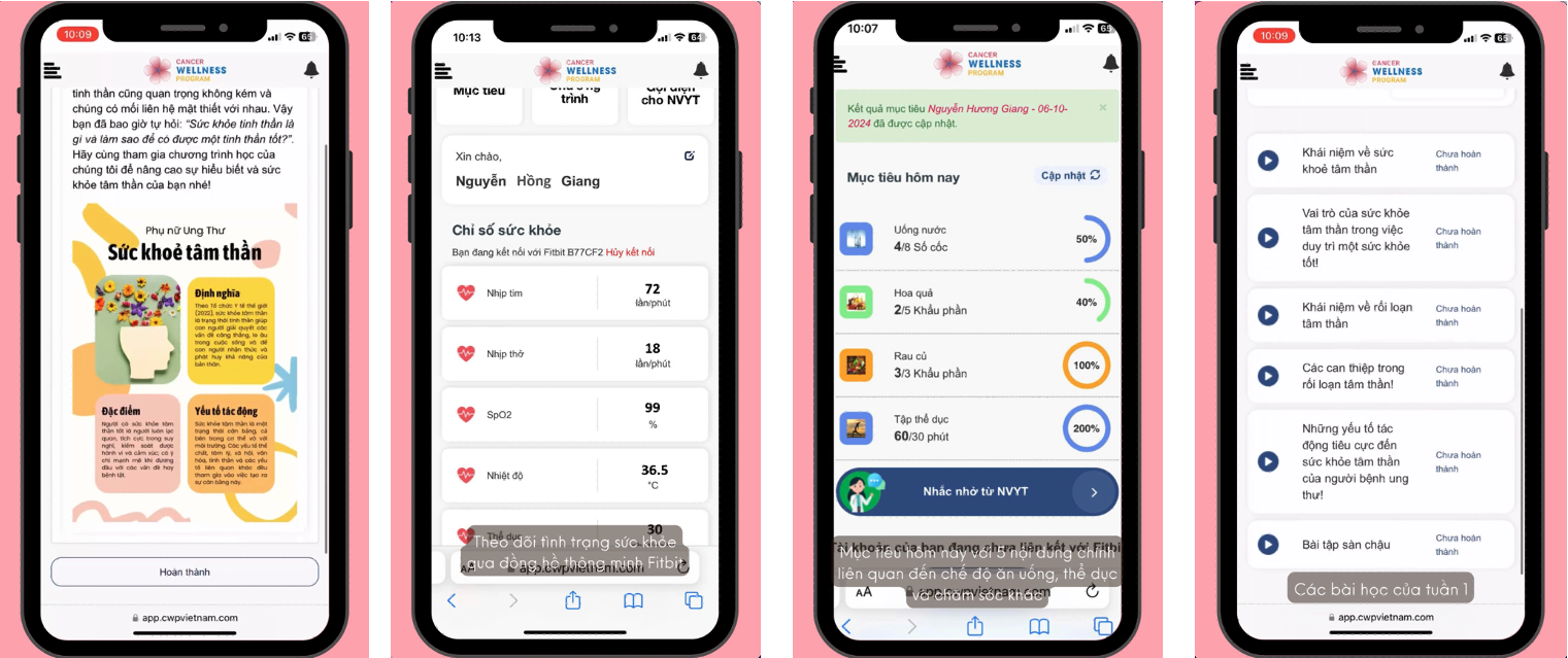
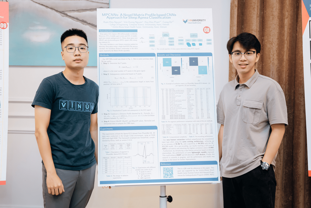
This area focuses on integrating artificial intelligence with biosensors to enhance the detection and analysis of biological signals. Applications include developing smart devices that can monitor health metrics in real-time, diagnose diseases early, and provide personalized health recommendations by analyzing data from sensors that detect biomarkers, physiological changes, and other biological indicators.
This research domain aims to digitize and analyze pathological data using advanced computational techniques. It involves creating and using digital slides and image analysis tools to identify disease patterns, quantify tissue characteristics, and assist pathologists in diagnosing diseases more accurately and efficiently, often leveraging machine learning and artificial intelligence.
This field combines multiple neuroimaging techniques, such as fMRI, PET, EEG, and MEG, to study the brain’s structure and function. By integrating data from different imaging modalities, researchers can gain a more comprehensive understanding of neural mechanisms, brain connectivity, and the relationship between brain activity and behavior, which is crucial for understanding neurological disorders and developing new treatments.
This area focuses on studying and quantifying human movement using technologies such as motion capture systems, wearable sensors, and computer vision. The goal is to analyze movement patterns to diagnose, monitor, and treat musculoskeletal and neurological conditions, improve rehabilitation techniques, and enhance the design of assistive devices by providing objective data on how patients move.
Latest News


Teaching
COMP3040: Computer Vision (Undergraduate)
COMP3040: Computer Vision (Undergraduate)
COMP1020: Object-Oriented Programming & Data Structures
BANA2030: Introduction to Programming with Python
NURS2220: Health Informatics
Collaborators